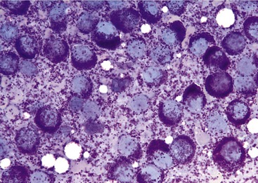
Qualified Mast Cell Tumor Cat Intestine Image
.It is found in humans and many animal species; Postmortem histopathology revealed an intestinal mast cell tumor.
Cutaneous mast cell tumors (cmcts) account for approximately 20% of skin neoplasms in cats.
Mast cells are a cell population that originate in the bone marrow and mature in connective tissue. Malignant mast cell tumors in cats usually involve the intestine or spleen, and the spleen can become dramatically enlarged. Compared to most abdominal tumors we diagnose it is a relatively rare form of tumor, and it is seen only in the cat. Mast cell tumors (mct) represent a commonly diagnosed cancer formed from specialized cells (mast cells) within the body that respond to inflammation and allergens. Cats with cutaneous mast cell tumors typically do effectively. Mast cell tumors can vary in appearance. Mast cells are present in most tissues, and are especially prominent in the skin, lining of the lungs and digestive tract, mouth and nose. Cutaneous mast cell tumors (cmcts) account for approximately 20% of skin neoplasms in cats. Mast cell tumors (mct) represent a cancer of mast cells, which are a type of white blood cell involved in allergy and inflammation. Very aggressive in both dogs and cats, high metastatic rate. There were more masses internally and this cat. What are mast cell tumors? What are mast cell tumors? Cats with cutaneous mast cell tumors typically do very well. The growths are not very likely to come back after surgery. Mast cell tumours are tumours which have arisen from mast cells. One tumor incompletely excised from the dermis, identified histologically without regional lymph node involvement? Mct in the small intestine often near the iliocolic junction. The pleomorphic tumors are more locally aggressive and harder to completely excise. What are the symptoms of mast cell tumors in cats? Mast cell tumors rarely affect the skin of cats, although it is the most common site for dogs. Tumor cells that separate from the margin of a tumor usually initially invade lymph vessels. Cats with a mast cell tumor in the spleen usually do far better than felines with a growth in the intestine. Most mcts are cured with appropriate local therapy, but a subset shows malignant behavior with the potential to spread to lymph nodes, liver, spleen, and other. A good prognostic indicator in the cat is appetite when first examined. Tumors are common in cats, especially as they get older. Ruby had a tumor in her small intestine called a mast cell tumor. It also can refer to an accumulation or nodule of mast cells that resembles a tumor. When this is the case, the tumor has often spread to other organs and the lymph nodes, which results in fluid in the cat's abdomen. The skin is the most common site for mast cell tumours in cats. Mast cell tumours commonly affect the skin, the spleen (in the abdomen) and/or the intestines.
